Mesothelioma
From Wikipedia, the free encyclopedia
Mesothelioma, more precisely
malignant mesothelioma, is a rare form of
cancer that develops from the protective lining that covers many of the body's internal organs, the
mesothelium. It is usually caused by exposure to
asbestos.
[1]
Its most common site is the
pleura (outer lining of the
lungs and internal chest wall), but it may also occur in the
peritoneum (the lining of the abdominal cavity), the
pericardium (a sac that surrounds the
heart),
[2] or the
tunica vaginalis (a sac that surrounds the testis).
Most people who develop mesothelioma have worked on jobs where they inhaled asbestos, or they have been exposed to asbestos dust and fiber in other ways. It has also been suggested that washing the clothes of a family member who worked with asbestos can put a person at risk for developing mesothelioma.
[3] Unlike lung cancer, there is no association between mesothelioma and
smoking, but smoking greatly increases the risk of other asbestos-induced cancers.
[4] Those who have been exposed to asbestos have collected damages for asbestos-related disease, including mesothelioma. Compensation via asbestos funds or lawsuits is an important issue in law practices regarding mesothelioma (see
asbestos and the law).
The symptoms of mesothelioma include
shortness of breath due to
pleural effusion (fluid between the lung and the
chest wall) or chest wall
pain, and general symptoms such as
weight loss. The diagnosis may be suspected with
chest X-ray and
CT scan, and is confirmed with a
biopsy (tissue sample) and
microscopic examination. A
thoracoscopy (inserting a tube with a camera into the chest) can be used to take biopsies. It allows the introduction of substances such as
talc to obliterate the pleural space (called
pleurodesis), which prevents more fluid from accumulating and pressing on the lung. Despite treatment with
chemotherapy,
radiation therapy or sometimes
surgery, the disease carries a poor
prognosis. Research about
screening tests for the early detection of mesothelioma is ongoing.
[edit] Signs and symptoms
Symptoms or signs of mesothelioma may not appear until 20 to 50 years (or more) after exposure to asbestos. Shortness of breath, cough, and pain in the chest due to an accumulation of fluid in the pleural space (
pleural effusion) are often symptoms of pleural mesothelioma.
Symptoms of
peritoneal mesothelioma include weight loss and
cachexia, abdominal swelling and pain due to
ascites (a buildup of fluid in the abdominal cavity). Other symptoms of
Peritoneal Mesothelioma may include bowel obstruction, blood clotting abnormalities,
anemia, and
fever. If the cancer has spread beyond the mesothelium to other parts of the body, symptoms may include pain, trouble swallowing, or swelling of the neck or face.
These symptoms may be caused by mesothelioma or by other, less serious conditions.
Mesothelioma that affects the pleura can cause these signs and symptoms:
- Chest wall pain
- Pleural effusion, or fluid surrounding the lung
- Shortness of breath
- Fatigue or anemia
- Wheezing, hoarseness, or cough
- Blood in the sputum (fluid) coughed up (hemoptysis)
In severe cases, the person may have many
tumor masses. The individual may develop a
pneumothorax, or collapse of the
lung. The disease may
metastasize, or spread, to other parts of the body.
Tumors that affect the abdominal cavity often do not cause symptoms until they are at a late stage. Symptoms include:
- Abdominal pain
- Ascites, or an abnormal buildup of fluid in the abdomen
- A mass in the abdomen
- Problems with bowel function
- Weight loss
In severe cases of the disease, the following signs and symptoms may be present:
- Blood clots in the veins, which may cause thrombophlebitis
- Disseminated intravascular coagulation, a disorder causing severe bleeding in many body organs
- Jaundice, or yellowing of the eyes and skin
- Low blood sugar level
- Pleural effusion
- Pulmonary emboli, or blood clots in the arteries of the lungs
- Severe ascites
A mesothelioma does not usually spread to the bone, brain, or adrenal glands. Pleural tumors are usually found only on one side of the lungs.
Working with
asbestos is the major risk factor for mesothelioma.
[5] In the United States, asbestos is the major cause of malignant mesothelioma and has been considered "indisputably"
[6] associated with the development of mesothelioma. Indeed, the relationship between asbestos and mesothelioma is so strong that many consider mesothelioma a “signal” or “sentinel” tumor.
[7][8][9][10] A history of asbestos exposure exists in most cases. However, mesothelioma has been reported in some individuals without any known exposure to asbestos. In rare cases, mesothelioma has also been associated with irradiation, intrapleural thorium dioxide (
Thorotrast), and inhalation of other fibrous silicates, such as
erionite. Some studies suggest that simian
virus 40 (
SV40) may act as a
cofactor in the development of mesothelioma.
[11]
Asbestos was known in antiquity, but it was not mined and widely used commercially until the late 19th century. Its use greatly increased during
World War II. Since the early 1940s, millions of American workers have been exposed to asbestos dust. Initially, the risks associated with asbestos exposure were not publicly known. However, an increased risk of developing mesothelioma was later found among shipyard workers, people who work in asbestos mines and mills, producers of asbestos products, workers in the heating and construction industries, and other tradespeople. Today, the official position of the U.S.
Occupational Safety and Health Administration (OSHA) and the U.S. EPA is that protections and "permissible exposure limits" required by U.S. regulations, while adequate to prevent most asbestos-related non-malignant disease, they are
not adequate to prevent or protect against asbestos-related cancers such as mesothelioma.
[12] Likewise, the British Government's
Health and Safety Executive (HSE) states formally that any threshold for mesothelioma must be at a very low level and it is widely agreed that if any such threshold does exist at all, then it cannot currently be quantified. For practical purposes, therefore, HSE assumes that no such "safe" threshold exists. Others have noted as well that there is no evidence of a threshold level below which there is no risk of mesothelioma.
[13] There appears to be a linear, dose-response relationship, with increasing dose producing increasing disease.
[14] Nevertheless, mesothelioma may be related to brief, low level or indirect exposures to asbestos.
[6] The dose necessary for effect appears to be lower for asbestos-induced mesothelioma than for pulmonary asbestosis or lung cancer.
[6] Again, there is no known safe level of exposure to asbestos as it relates to increased risk of mesothelioma.
The duration of exposure to asbestos causing mesothelioma can be short. For example, cases of mesothelioma have been documented with only 1–3 months of exposure.
[15][16] People who work with asbestos wear personal protective equipment to lower their risk of exposure.
Latency, the time from first exposure to manifestation of disease, is prolonged in the case of mesothelioma. It is virtually never less than fifteen years and peaks at 30–40 years.
[6] In a review of occupationally related mesothelioma cases, the median latency was 32 years.
[17] Based upon the data from Peto
et al., the risk of mesothelioma appears to increase to the third or fourth power from first exposure.
[14]
[edit] Environmental exposures
Incidence of mesothelioma had been found to be higher in populations living near naturally occurring asbestos. For example, in central
Cappadocia, Turkey, mesothelioma was causing 50% of all deaths in three small villages — Tuzköy, Karain and Sarıhıdır. Initially, this was attributed to
erionite, a
zeolite mineral with similar properties to
asbestos. Recently, however, detailed epidemiological investigation showed that erionite causes mesothelioma mostly in families with a genetic predisposition.
[18][19] The documented presence of asbestos fibers in water supplies and food products has fostered concerns about the possible impact of long-term and, as yet, unknown exposure of the general population to these fibers.
[edit] Occupational
Exposure to asbestos fibers has been recognized as an occupational health hazard since the early 20th century. Numerous epidemiological studies have associated occupational exposure to asbestos with the development of pleural plaques, diffuse pleural thickening, asbestosis, carcinoma of the lung and larynx, gastrointestinal tumors, and diffuse malignant mesothelioma of the pleura and peritoneum. Asbestos has been widely used in many industrial products, including cement, brake linings, gaskets, roof shingles, flooring products, textiles, and insulation.
Commercial asbestos mining at Wittenoom, Western Australia, occurred between 1945 and 1966. A cohort study of miners employed at the mine reported that while no deaths occurred within the first 10 years after
crocidolite exposure, 85 deaths attributable to mesothelioma had occurred by 1985. By 1994, 539 reported deaths due to mesothelioma had been reported in Western Australia.
[edit] Paraoccupational secondary exposure
Family members and others living with asbestos workers have an increased risk of developing mesothelioma, and possibly other asbestos related diseases.
[20][21] This risk may be the result of exposure to asbestos dust brought home on the clothing and hair of asbestos workers. To reduce the chance of exposing family members to asbestos fibres, asbestos workers are usually required to shower and change their clothing before leaving the workplace.
[edit] Asbestos in buildings
Many building materials used in both public and domestic premises prior to the banning of asbestos may contain asbestos. Those performing renovation works or
DIY activities may expose themselves to asbestos dust. In the UK use of Chrysotile asbestos was banned at the end of 1999. Brown and
blue asbestos was banned in the UK around 1985. Buildings built or renovated prior to these dates may contain asbestos materials.
[edit] Diagnosis

CXR demonstrating a mesothelioma
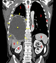
CT scan of a patient with mesothelioma,
coronal section (the section follows the plane that divides the body in a front and a back half). The mesothelioma is indicated by yellow arrows, the central
pleural effusion (fluid collection) is marked with a yellow star. Red numbers: (1) right lung, (2) spine, (3) left lung, (4) ribs, (5)
descending part of the
aorta, (6)
spleen, (7) left
kidney, (8) right kidney, (9)
liver.
Diagnosing mesothelioma is often difficult, because the symptoms are similar to those of a number of other conditions. Diagnosis begins with a review of the patient's medical history. A history of exposure to asbestos may increase clinical suspicion for mesothelioma. A physical examination is performed, followed by
chest X-ray and often
lung function tests. The X-ray may reveal pleural thickening commonly seen after asbestos exposure and increases suspicion of mesothelioma. A
CT (or CAT) scan or an
MRI is usually performed. If a large amount of fluid is present, abnormal cells may be detected by
cytopathology if this fluid is
aspirated with a syringe. For pleural fluid, this is done by
thoracentesis or tube thoracostomy (
chest tube); for ascites, with
paracentesis or
ascitic drain; and for
pericardial[disambiguation needed  ]
] effusion with
pericardiocentesis. While absence of malignant cells on cytology does not completely exclude mesothelioma, it makes it much more unlikely, especially if an alternative diagnosis can be made (e.g.
tuberculosis,
heart failure). Unfortunately, the diagnosis of malignant mesothelioma by cytology alone is difficult, even with expert pathologists.
Generally, a
biopsy is needed to confirm a diagnosis of malignant mesothelioma. A doctor removes a sample of tissue for examination under a microscope by a
pathologist. A biopsy may be done in different ways, depending on where the abnormal area is located. If the cancer is in the chest, the doctor may perform a
thoracoscopy. In this procedure, the doctor makes a small cut through the chest wall and puts a thin, lighted tube called a thoracoscope into the chest between two ribs. Thoracoscopy allows the doctor to look inside the chest and obtain tissue samples. Alternatively, the chest surgeon might directly open the chest (
thoracotomy). If the cancer is in the abdomen, the doctor may perform a
laparoscopy. To obtain tissue for examination, the doctor makes a small incision in the abdomen and inserts a special instrument into the abdominal cavity. If these procedures do not yield enough tissue, more extensive diagnostic surgery may be necessary.
Immunohistochemical studies play an important role for the pathologist in differentiating malignant mesothelioma from neoplastic mimics. There are numerous tests and panels available. No single test is perfect for distinguishing mesothelioma from carcinoma or even benign versus malignant.
There are three histological types of malignant mesothelioma: (1) Epithelioid; (2) Sarcomatoid; and (3) Biphasic (Mixed). Epithelioid comprises about 50-60% of malignant mesothelioma cases and generally holds a better prognosis than the Sarcomatoid or Biphasic subtypes.
[22]
[edit] Staging
Staging of mesothelioma is based on the recommendation by the International Mesothelioma Interest Group.
[23] TNM classification of the primary tumor,
lymph node involvement, and distant
metastasis is performed. Mesothelioma is staged Ia–IV (one-A to four) based on the TNM status.
[23][24]
[edit] Screening
There is no universally agreed protocol for screening people who have been exposed to asbestos. Screening tests might diagnose mesothelioma earlier than conventional methods thus improving the survival prospects for patients. The
serum osteopontin level might be useful in screening asbestos-exposed people for mesothelioma. The level of soluble mesothelin-related protein is elevated in the serum of about 75% of patients at diagnosis and it has been suggested that it may be useful for screening.
[25] Doctors have begun testing the
Mesomark assay which measures levels of soluble
mesothelin-related proteins (SMRPs) released by diseased mesothelioma cells.
[26]
[edit] Pathophysiology
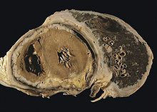
Diffuse pleural mesothelioma with extensive involvement of the pericardium.
The
mesothelium consists of a single layer of flattened to cuboidal cells forming the
epithelial lining of the serous cavities of the body including the
peritoneal,
pericardial and
pleural cavities. Deposition of asbestos fibers in the
parenchyma of the lung may result in the penetration of the
visceral pleura from where the fiber can then be carried to the pleural surface, thus leading to the development of malignant mesothelial plaques. The processes leading to the development of
peritoneal mesothelioma remain unresolved, although it has been proposed that asbestos fibers from the lung are transported to the abdomen and associated organs via the
lymphatic system. Additionally, asbestos fibers may be deposited in the gut after ingestion of sputum contaminated with asbestos fibers.
Pleural contamination with asbestos or other mineral fibers has been shown to cause cancer. Long thin asbestos fibers (blue asbestos,
amphibole fibers) are more potent
carcinogens than "feathery fibers" (
chrysotile or white asbestos fibers).
[6] However, there is now evidence that smaller particles may be more dangerous than the larger fibers. They remain suspended in the air where they can be inhaled, and may penetrate more easily and deeper into the lungs. "We probably will find out a lot more about the health aspects of asbestos from [the World Trade Center attack], unfortunately," said Dr. Alan Fein, chief of pulmonary and critical-care medicine at North Shore-Long Island Jewish Health System. Dr. Fein has treated several patients for "World Trade Center syndrome" or respiratory ailments from brief exposures of only a day or two near the collapsed buildings.
[27]
Mesothelioma development in rats has been demonstrated following intra-pleural inoculation of phosphorylated chrysotile fibers. It has been suggested that in humans, transport of fibers to the pleura is critical to the pathogenesis of mesothelioma. This is supported by the observed recruitment of significant numbers of
macrophages and other cells of the
immune system to localized lesions of accumulated asbestos fibers in the pleural and peritoneal cavities of rats. These lesions continued to attract and accumulate macrophages as the disease progressed, and cellular changes within the lesion culminated in a morphologically malignant tumor.
Experimental evidence suggests that asbestos acts as a complete carcinogen with the development of mesothelioma occurring in sequential stages of initiation and promotion. The molecular mechanisms underlying the malignant transformation of normal mesothelial cells by asbestos fibers remain unclear despite the demonstration of its oncogenic capabilities (see next-but-one paragraph). However, complete in vitro transformation of normal human mesothelial cells to malignant
phenotype following exposure to asbestos fibers has not yet been achieved. In general, asbestos fibers are thought to act through direct physical interactions with the cells of the mesothelium in conjunction with indirect effects following interaction with inflammatory cells such as macrophages.
Analysis of the interactions between asbestos fibers and
DNA has shown that phagocytosed fibers are able to make contact with
chromosomes, often adhering to the
chromatin fibers or becoming entangled within the chromosome. This contact between the asbestos fiber and the chromosomes or structural proteins of the spindle apparatus can induce complex abnormalities. The most common abnormality is
monosomy of chromosome 22. Other frequent abnormalities include structural rearrangement of 1p, 3p, 9p and 6q chromosome arms.
Common gene abnormalities in mesothelioma cell lines include deletion of the
tumor suppressor genes:
Asbestos has also been shown to mediate the entry of foreign DNA into target cells. Incorporation of this foreign DNA may lead to mutations and oncogenesis by several possible mechanisms:
Asbestos fibers have been shown to alter the function and secretory properties of macrophages, ultimately creating conditions which favour the development of mesothelioma. Following asbestos phagocytosis, macrophages generate increased amounts of hydroxyl
radicals, which are normal by-products of cellular anaerobic metabolism. However, these free radicals are also known
clastogenic and membrane-active agents thought to promote asbestos carcinogenicity. These oxidants can participate in the oncogenic process by directly and indirectly interacting with DNA, modifying membrane-associated cellular events, including oncogene activation and perturbation of cellular antioxidant defences.
Asbestos also may possess
immunosuppressive properties. For example, chrysotile fibres have been shown to depress the in vitro proliferation of phytohemagglutinin-stimulated peripheral blood lymphocytes, suppress natural killer cell lysis and significantly reduce
lymphokine-activated killer cell viability and recovery. Furthermore, genetic alterations in asbestos-activated macrophages may result in the release of potent mesothelial cell mitogens such as
platelet-derived growth factor (PDGF) and
transforming growth factor-β (TGF-β) which in turn, may induce the chronic stimulation and proliferation of mesothelial cells after injury by asbestos fibres.
[edit] Treatment
The prognosis for malignant mesothelioma remains disappointing, although there have been some modest improvements in prognosis from newer chemotherapies and multimodality treatments.
[28] Treatment of malignant mesothelioma at earlier stages has a better prognosis, but cures are exceedingly rare. Clinical behavior of the malignancy is affected by several factors including the continuous mesothelial surface of the pleural cavity which favors local metastasis via exfoliated cells, invasion to underlying tissue and other organs within the pleural cavity, and the extremely long latency period between asbestos exposure and development of the disease. The histological subtype and the patient's age and health status also help predict prognosis. The epithelioid histology responds better to treatment and has a survival advantage over sarcomatoid histology.
[29]
[edit] Surgery
Surgery, by itself, has proved disappointing. In one large series, the median survival with surgery (including extrapleural
pneumonectomy) was only 11.7 months.
[28] However, research indicates varied success when used in combination with radiation and chemotherapy (Duke, 2008). (For more information on multimodality therapy with surgery, see below). A pleurectomy/decortication is the most common surgery, in which the lining of the chest is removed. Less common is an extrapleural pneumonectomy (EPP), in which the lung, lining of the inside of the chest, the hemi-
diaphragm and the
pericardium are removed.
[edit] Radiation
For patients with localized disease, and who can tolerate a radical surgery, radiation is often given post-operatively as a consolidative treatment. The entire hemi-thorax is treated with radiation therapy, often given simultaneously with chemotherapy. Delivering radiation and chemotherapy after a radical surgery has led to extended life expectancy in selected patient populations with some patients surviving more than 5 years. As part of a curative approach to mesothelioma, radiotherapy is also commonly applied to the sites of
chest drain insertion, in order to prevent growth of the tumor along the track in the chest wall.
Although mesothelioma is generally resistant to curative treatment with
radiotherapy alone, palliative treatment regimens are sometimes used to relieve symptoms arising from tumor growth, such as obstruction of a major blood vessel. Radiation therapy when given alone with curative intent has never been shown to improve survival from mesothelioma. The necessary radiation dose to treat mesothelioma that has not been surgically removed would be very toxic.
[edit] Chemotherapy
Chemotherapy is the only treatment for mesothelioma that has been proven to improve survival in randomised and controlled trials. The landmark study published in 2003 by Vogelzang and colleagues compared
cisplatin chemotherapy alone with a combination of cisplatin and
pemetrexed (brand name Alimta) chemotherapy in patients who had not received chemotherapy for malignant pleural mesothelioma previously and were not candidates for more aggressive "curative" surgery.
[30] This trial was the first to report a survival advantage from chemotherapy in malignant pleural mesothelioma, showing a statistically significant improvement in
median survival from 10 months in the patients treated with cisplatin alone to 13.3 months in the group of patients treated with cisplatin in the combination with pemetrexed and who also received supplementation with folate and vitamin B
12. Vitamin supplementation was given to most patients in the trial and pemetrexed related side effects were significantly less in patients receiving pemetrexed when they also received daily oral folate 500mcg and intramuscular vitamin B
12 1000mcg every 9 weeks compared with patients receiving pemetrexed without vitamin supplementation. The objective response rate increased from 20% in the cisplatin group to 46% in the combination pemetrexed group. Some side effects such as nausea and vomiting,
stomatitis, and diarrhoea were more common in the combination pemetrexed group but only affected a minority of patients and overall the combination of pemetrexed and cisplatin was well tolerated when patients received vitamin supplementation; both
quality of life and
lung function tests improved in the combination pemetrexed group. In February 2004, the United States
Food and Drug Administration approved pemetrexed for treatment of malignant pleural mesothelioma. However, there are still unanswered questions about the optimal use of chemotherapy, including when to start treatment, and the optimal number of cycles to give.
Cisplatin in combination with
raltitrexed has shown an improvement in survival similar to that reported for pemetrexed in combination with cisplatin, but raltitrexed is no longer commercially available for this indication. For patients unable to tolerate pemetrexed, cisplatin in combination with gemcitabine or vinorelbine is an alternative, or vinorelbine on its own, although a survival benefit has not been shown for these drugs. For patients in whom cisplatin cannot be used, carboplatin can be substituted but non-randomised data have shown lower response rates and high rates of haematological toxicity for carboplatin-based combinations, albeit with similar survival figures to patients receiving cisplatin.
[31]
In January 2009, the United States FDA approved using conventional therapies such as surgery in combination with radiation and or chemotherapy on stage I or II Mesothelioma after research conducted by a nationwide study by Duke University concluded an almost 50 point increase in remission rates.
[edit] Immunotherapy
Treatment regimens involving immunotherapy have yielded variable results. For example, intrapleural inoculation of
Bacillus Calmette-Guérin (BCG) in an attempt to boost the immune response, was found to be of no benefit to the patient (while it may benefit patients with
bladder cancer). Mesothelioma cells proved susceptible to in vitro lysis by LAK cells following activation by
interleukin-2 (IL-2), but patients undergoing this particular therapy experienced major side effects. Indeed, this trial was suspended in view of the unacceptably high levels of IL-2 toxicity and the severity of side effects such as fever and cachexia. Nonetheless, other trials involving interferon alpha have proved more encouraging with 20% of patients experiencing a greater than 50% reduction in tumor mass combined with minimal side effects.
[edit] Heated Intraoperative Intraperitoneal Chemotherapy
A procedure known as heated intraoperative intraperitoneal chemotherapy was developed by Paul Sugarbaker at the Washington Cancer Institute.
[32] The surgeon removes as much of the tumor as possible followed by the direct administration of a chemotherapy agent, heated to between 40 and 48°C, in the abdomen. The fluid is perfused for 60 to 120 minutes and then drained.
This technique permits the administration of high concentrations of selected drugs into the abdominal and pelvic surfaces. Heating the chemotherapy treatment increases the penetration of the drugs into tissues. Also, heating itself damages the malignant cells more than the normal cells.
This technique is also used in patients with malignant pleural mesothelioma.
[33]
[edit] Multimodality Therapy
All of the standard approaches to treating solid tumors—radiation, chemotherapy, and surgery—have been investigated in patients with malignant pleural mesothelioma. Although surgery, by itself, is not very effective, surgery combined with adjuvant chemotherapy and radiation (trimodality therapy) has produced significant survival extension (3–14 years) among patients with favorable prognostic factors.
[34] However, other large series of examining multimodality treatment have only demonstrated modest improvement in survival (median survival 14.5 months and only 29.6% surviving 2 years).
[28] Reducing the bulk of the tumor with cytoreductive surgery is key to extending survival. Two surgeries have been developed: extrapleural pneumonectomy and pleurectomy/decortication. The indications for performing these operations are unique. The choice of operation depends on the size of the patient's tumor. This is an important consideration because tumor volume has been identified as a prognostic factor in mesothelioma.
[35] Pleurectomy/decortication spares the underlying lung and is performed in patients with early stage disease when the intention is to remove all gross visible tumor (macroscopic complete resection), not simply palliation.
[36] Extrapleural pneumonectomy is a more extensive operation that involves resection of the parietal and visceral pleurae, underlying lung, ipsilateral diaphragm, and ipsilateral pericardium. This operation is indicated for a subset of patients with more advanced tumors, who can tolerate a pneumonectomy.
[37]
[edit] Epidemiology
Although reported incidence rates have increased in the past 20 years, mesothelioma is still a relatively rare cancer. The incidence rate varies from one country to another, from a low rate of less than 1 per 1,000,000 in Tunisia and Morocco, to the highest rate in Britain, Australia and Belgium: 30 per 1,000,000 per year.
[38] For comparison, populations with high levels of smoking can have a
lung cancer incidence of over 1,000 per 1,000,000. Incidence of malignant mesothelioma currently ranges from about 7 to 40 per 1,000,000 in industrialized Western nations, depending on the amount of asbestos exposure of the populations during the past several decades.
[39] It has been estimated that incidence may have peaked at 15 per 1,000,000 in the United States in 2004. Incidence is expected to continue increasing in other parts of the world. Mesothelioma occurs more often in men than in women and risk increases with age, but this disease can appear in either men or women at any age. Approximately one fifth to one third of all mesotheliomas are peritoneal.
Between 1940 and 1979, approximately 27.5 million people were occupationally exposed to asbestos in the United States.
[40] Between 1973 and 1984, the incidence of pleural mesothelioma among Caucasian males increased 300%. From 1980 to the late 1990s, the death rate from mesothelioma in the USA increased from 2,000 per year to 3,000, with men four times more likely to acquire it than women. These rates may not be accurate, since it is possible that many cases of mesothelioma are misdiagnosed as adenocarcinoma of the lung, which is difficult to differentiate from mesothelioma.
Source : http://en.wikipedia.org/wiki/Mesothelioma


